The Anatomy of The Knee
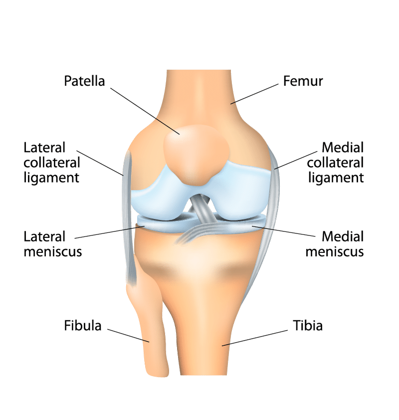
The knee is the largest joint in the body. Your knee helps you perform most everyday activities. The knee is made up of the lower end of the thigh bone (femur) and the shin bone (tibia) to bend and straighten the lower leg. The kneecap (patella) slides in a groove on the end of the femur. Large ligaments attach to the femur and tibia to provide stability. The long thigh muscles (quadriceps) give the knee strength.
The joint surfaces where these three bones touch are covered with articular cartilage, which is a smooth substance that cushions the bones and helps them move easily. The medial and lateral meniscus are crescent-shaped bands of thick rubbery cartilage attached to the shin bone (tibia), that act as shock absorbers and stabilize the knee.
All other surfaces of the knee are covered by a thin, smooth tissue liner called the synovial membrane. This membrane releases a special fluid that lubricates the knee, reducing friction to nearly zero in a healthy knee.
Normally, all these parts work in harmony, but disease or injury can disrupt this harmony, resulting in pain, muscle weakness and reduced function.